في إنجاز طبي وصف بأنه الأول من نوعه عالميًا، نجح جراحون بريطانيون في استئصال ورم من دماغ مريض باستخدام نظام للذكاء الاصطناعي قدّم لهم إرشادات مباشرة أثناء إجراء العملية، وساعدهم على تحديد مواقع الأوعية الدموية والأعصاب الحيوية وتجنبها.
وبحسب هيئة الإذاعة البريطانية BBC، خضع البريطاني ريس هيبرت، البالغ من العمر 48 عامًا والأب لطفلين من مقاطعة بيدفوردشير، للجراحة في مستشفى (يونيفرسيتي كوليدج لندن)، بعدما بدأ ورم حميد ينمو بالقرب من الغدة النخامية ويضغط على الأعصاب البصرية، مهددًا بفقدان جزء من مجال رؤيته، وفقًا للجراحين.
ورم صغير في موقع شديد الحساسية
وقبل نحو 18 شهرًا، كان هيبرت يتمتع بصحة جيدة ويمارس حياته بصورة طبيعية، وكان يقطع نحو ميلين سيرًا على الأقدام خلال فترة الغداء يوميًا. لكن خلال إحدى جولاته سقط فاقدًا الوعي وتعرض لنوبة تشنج في الشارع.
وأظهر التصوير الطبي وجود ورم غير سرطاني يبلغ حجمه 11 ملليمترًا، ينمو في الغدة النخامية، وهي غدة صغيرة تقع في قاعدة الجمجمة وتؤدي دورًا أساسيًا في إفراز الهرمونات المنظمة لعدد من وظائف الجسم؛ من بينها النمو والتمثيل الغذائي وضغط الدم ودرجة حرارة الجسم.
وتكمن صعوبة استئصال الورم في موقعه القريب للغاية من الشرايين السباتية، التي تمد الدماغ بالدم، ومن الأعصاب البصرية المسؤولة عن الرؤية.
ومع بدء الورم في الضغط على الأعصاب البصرية، ضاق مجال الرؤية الطرفية لدى هيبرت، كما عانى من إرهاق شديد ودوخة وأصبح يتعثر بالأشياء أثناء المشي.
وقال هيبرت: إن التأثير الأكبر للمرض كان نفسيًا وعاطفيًا، مشيرًا إلى أنه لم يكن يريد أن يصبح عبئًا على الآخرين، وأضاف أن حرصه على المشاركة في التجربة البحثية نابع من إيمانه بأن تقدم الطب يعتمد على استعداد المرضى للمشاركة في الدراسات والتجارب الجديدة.
«عين ثانية» داخل غرفة العمليات
واعتمدت العملية على إدخال منظار رفيع مزود بكاميرا عبر الأنف وصولًا إلى قاعدة الجمجمة، حيث كان الورم موجودًا خلف طبقات من العظام وغشاء قوي، ما جعل تحديد المسار الأكثر أمانًا للوصول إليه أمرًا معقدًا، خصوصًا مع اختلاف تشريح الدماغ من شخص إلى آخر.
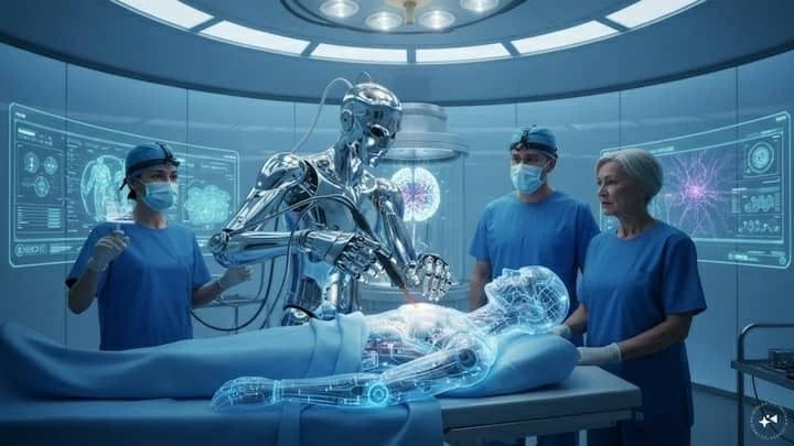
وهنا لعب نظام الذكاء الاصطناعي دوره، فقد عرض الجراحون بثًا مباشرًا للعملية على شاشة ثانية، بينما قام النظام بتحليل الصور في الوقت الفعلي، وتتبع الأدوات الجراحية، وحدد المناطق التي يُرجح وجود الأوعية الدموية والأعصاب الحيوية فيها، مع إبراز المواضع التي يمكن للجراحين من خلالها إزالة الورم بأكبر قدر ممكن من الأمان.
ويشبه النظام، من حيث المبدأ، تقنيات التعرف على الوجوه المستخدمة في الهواتف الذكية، لكنه بدلًا من التعرف على ملامح الأشخاص، جرى تدريبه على التعرف على تراكيب تشريحية مهمة، بينها الأوعية والأعصاب التي قد تكون مخفية عن رؤية الجراح.
وقال البروفيسور هاني ماركوس، أحد المشاركين في إجراء العملية: إن النظام تدرب على عدد من العمليات يفوق ما قد يشاهده أو يجريه معظم الجراحين خلال حياتهم المهنية، ويمكنه أن يعمل بمثابة «زوج ثانٍ من العيون» يمتلك خبرة واسعة.
وأكد ماركوس أن أبرز ما يميز هذه التقنية التجريبية هو قدرتها على العمل في الوقت الفعلي أثناء الجراحة، وليس اعتمادًا على تحليل الصور بعد انتهاء العملية.
الجراح يحتفظ بالقرار النهائي
وشدد الفريق الطبي على أن الذكاء الاصطناعي لا يحل محل الجراح، بل يعمل كأداة مساعدة تقدم المعلومات والتوجيهات، بينما تظل السيطرة الكاملة والقرار النهائي في يد الفريق الجراحي.
ويأمل الباحثون في تطوير النظام مستقبلًا ليصبح أشبه بـ(ChatGPT للجراحين)، بحيث يستطيع الطبيب الاستعانة بخبير رقمي إضافي داخل غرفة العمليات عند الحاجة، مع إمكانية تجاهل توصياته إذا رأى الجراح أنها غير مناسبة.
وخضع النظام لسنوات من الاختبارات والتدريب في المختبر، حيث استخدم الباحثون مئات التسجيلات المصورة لعمليات استئصال أورام الغدة النخامية، وقاموا بتحديد مواقع الأوعية والأعصاب يدويًا في التسجيلات لتزويد النظام بالبيانات اللازمة لتعلم الأنماط التشريحية المهمة.
مخاطر معقدة وفرصة أكبر للاستئصال الكامل
وكان الأطباء يواجهون قبل استخدام هذه التقنية احتمالات معقدة أثناء استئصال هذا النوع من الأورام؛ إذ تراوحت فرصة عدم إزالة الورم بالكامل بين 25 و50%، بينما تراوحت احتمالية إصابة وعاء دموي رئيسي بين 0.5 و2%.
ويرى الباحثون أن قدرة الذكاء الاصطناعي على تحديد التراكيب الحيوية أثناء الجراحة قد تساعد في تحسين مستوى الأمان، وتمكين الجراح من إزالة أكبر قدر ممكن من الورم دون الإضرار بالأوعية أو الأعصاب الحساسة.
ويجري تمويل المشروع من المعهد الوطني البريطاني لأبحاث الصحة والرعاية، إلى جانب دعم من شركة «جوجل»، فيما يستعد الفريق لإجراء تجربة سريرية أكبر لتقييم التقنية على عدد أكبر من المرضى.
«أعاد إليّ حياتي»
وبعد العملية، قال هيبرت: إنه شعر بتحسن كبير في الرؤية فور استعادة وعيه، مشيرًا إلى أنه شعر وكأن مجال رؤيته أصبح واسعًا بصورة لم يعهدها منذ نحو عام.
وبعد مرور ثمانية أسابيع على الجراحة، أكد أن بصره واصل التحسن بصورة ملحوظة، كما عادت إليه طاقته واختفت الأعراض الجانبية التي كان يعاني منها قبل العملية، وقال هيبرت: «لقد أعادت إليّ حياتي».
من جانبه، أعرب وزير الابتكار الصحي البريطاني جيمس فريث عن سعادته بخضوع هيبرت لهذه الجراحة التي وصفها بأنها «غيّرت حياته»، باعتبارها جزءًا من أبحاث ممولة حكوميًا.
وأكد أن استخدام الذكاء الاصطناعي في الرعاية الصحية يجب أن يخضع لضمانات وضوابط صارمة تضع سلامة المرضى في المقام الأول، مع الاستفادة في الوقت نفسه من الفرص التي تتيحها هذه التكنولوجيا لتطوير الطب وتحسين نتائج العلاج.
ويمثل نجاح هذه العملية خطوة لافتة في اتجاه دمج الذكاء الاصطناعي مع جراحات المخ والأعصاب، ليس باعتباره بديلًا عن الطبيب، وإنما كأداة مساعدة قادرة على تحليل المعلومات البصرية المعقدة لحظة بلحظة وتقديم «عين ثانية» للجراح في واحدة من أكثر البيئات الطبية دقة وحساسية.
In a medical achievement described as a world first, British surgeons successfully removed a tumor from a patient's brain using an artificial intelligence system that provided them with real-time guidance during the operation, helping them identify and avoid vital blood vessels and nerves.
According to the BBC, 48-year-old Rhys Hibbert, a father of two from Bedfordshire, underwent surgery at University College London Hospital after a benign tumor began to grow near his pituitary gland, pressing on his optic nerves and threatening to impair part of his vision, according to the surgeons.
A Small Tumor in a Highly Sensitive Location
About 18 months ago, Hibbert was in good health and living his life normally, walking about two miles during his lunch break each day. However, during one of his walks, he collapsed, lost consciousness, and suffered a seizure in the street.
Medical imaging revealed a non-cancerous tumor measuring 11 millimeters, growing in the pituitary gland, a small gland located at the base of the skull that plays a crucial role in releasing hormones that regulate various bodily functions, including growth, metabolism, blood pressure, and body temperature.
The difficulty in removing the tumor lies in its proximity to the carotid arteries, which supply blood to the brain, and the optic nerves responsible for vision.
As the tumor began to press on the optic nerves, Hibbert's peripheral vision narrowed, and he experienced severe fatigue and dizziness, often stumbling over objects while walking.
Hibbert stated that the most significant impact of the illness was psychological and emotional, noting that he did not want to become a burden on others. He added that his eagerness to participate in the research trial stemmed from his belief that medical advancement relies on patients' willingness to engage in new studies and trials.
A "Second Eye" in the Operating Room
The operation involved inserting a thin endoscope equipped with a camera through the nose to the base of the skull, where the tumor was located behind layers of bone and a tough membrane, making it complex to determine the safest path to reach it, especially given the variations in brain anatomy from person to person.
Here, the artificial intelligence system played its role, as the surgeons displayed a live feed of the operation on a second screen while the system analyzed images in real-time, tracked surgical instruments, and identified areas likely containing vital blood vessels and nerves, highlighting the spots from which the surgeons could safely remove the tumor.
The system is similar, in principle, to facial recognition technologies used in smartphones, but instead of recognizing people's features, it has been trained to identify important anatomical structures, including vessels and nerves that may be hidden from the surgeon's view.
Professor Hani Marcus, one of the participants in the operation, stated that the system has trained on a number of procedures exceeding what most surgeons would see or perform in their careers, and it can act as a "second pair of eyes" with extensive experience.
Marcus emphasized that the most notable aspect of this experimental technique is its ability to operate in real-time during surgery, rather than relying on image analysis after the procedure is completed.
The Surgeon Retains the Final Decision
The medical team stressed that artificial intelligence does not replace the surgeon but acts as an assisting tool that provides information and guidance, while full control and the final decision remain in the hands of the surgical team.
Researchers hope to develop the system in the future to resemble a "ChatGPT for surgeons," allowing doctors to consult a digital expert in the operating room when needed, with the option to disregard its recommendations if the surgeon deems them inappropriate.
The system underwent years of testing and training in the lab, where researchers used hundreds of video recordings of pituitary tumor removal surgeries and manually identified the locations of vessels and nerves in the recordings to provide the system with the necessary data to learn important anatomical patterns.
Complex Risks and Greater Opportunity for Complete Removal
Before using this technology, doctors faced complex probabilities during the removal of this type of tumor; the chance of not completely removing the tumor ranged from 25% to 50%, while the likelihood of injuring a major blood vessel ranged from 0.5% to 2%.
Researchers believe that the ability of artificial intelligence to identify vital structures during surgery may help improve safety levels, enabling the surgeon to remove as much of the tumor as possible without damaging sensitive vessels or nerves.
The project is funded by the UK National Institute for Health Research, with support from Google, as the team prepares to conduct a larger clinical trial to assess the technique on a greater number of patients.
"It Gave Me My Life Back"
After the operation, Hibbert stated that he felt a significant improvement in his vision as soon as he regained consciousness, noting that it felt as if his field of vision had widened in a way he hadn't experienced for about a year.
Eight weeks after the surgery, he confirmed that his eyesight continued to improve significantly, his energy returned, and the side effects he had experienced before the operation disappeared. Hibbert said, "It gave me my life back."
For his part, British Health Innovation Minister James Freeth expressed his happiness that Hibbert underwent this surgery, which he described as "life-changing," considering it part of government-funded research.
He emphasized that the use of artificial intelligence in healthcare must be subject to strict guarantees and controls that prioritize patient safety while simultaneously leveraging the opportunities this technology offers to advance medicine and improve treatment outcomes.
The success of this operation represents a significant step towards integrating artificial intelligence with neurosurgery, not as a replacement for the doctor, but as an assisting tool capable of analyzing complex visual information moment by moment and providing a "second eye" for the surgeon in one of the most precise and sensitive medical environments.